Interaction Vertex Imaging
Experimental setup at the National Superconducting Cyclotron Laboratory, Michigan, USA. Two silicon detectors were used here to determine the range of an O-16 beam with high accuracy.
Experimental setup at the National Superconducting Cyclotron Laboratory, Michigan, USA. Two silicon detectors were used here to determine the range of an O-16 beam with high accuracy.

Patients normally sit here. This time, Dr. Cornelia Höhr (Deputy Director - Life Sciences at TRIUMF) and PhD student Eva Kasanda (University of Guelph) are testing a method for range verification by detecting gamma radiation.

The Hadron Tumor Marker is a small metallic implant. The beam reacts with the implant and emits characteristic radiation. This radiation can be used to determine exactly where the beam is located in the patient.

Semiconductor detectors are used to detect particles leaving the patient during irradiation with carbon ions. The track of the particles can be used to calculate where the beam is located in the patient.

The TRIUMF research center is home to one of the largest cyclotron accelerators in the world. The radiation room for patients is located directly next to the accelerator. Many patients with eye tumors have been irradiated here in recent decades.
Many modern medical methods have developed from applications of nuclear physics. Modern imaging techniques such as magnetic resonance imaging, positron emission tomography or heart examinations using radioactive contrast agents are just as much examples as radiation therapy for cancer patients.
Our research group is actively involved in various cancer research projects, both locally at the University of Cologne and within international collaborations.
One research focus is radiotherapy using protons and carbon ions. Radiotherapy is an important pillar of cancer therapy, and almost half of cancer patients receive radiotherapy, often in combination with other forms of therapy.
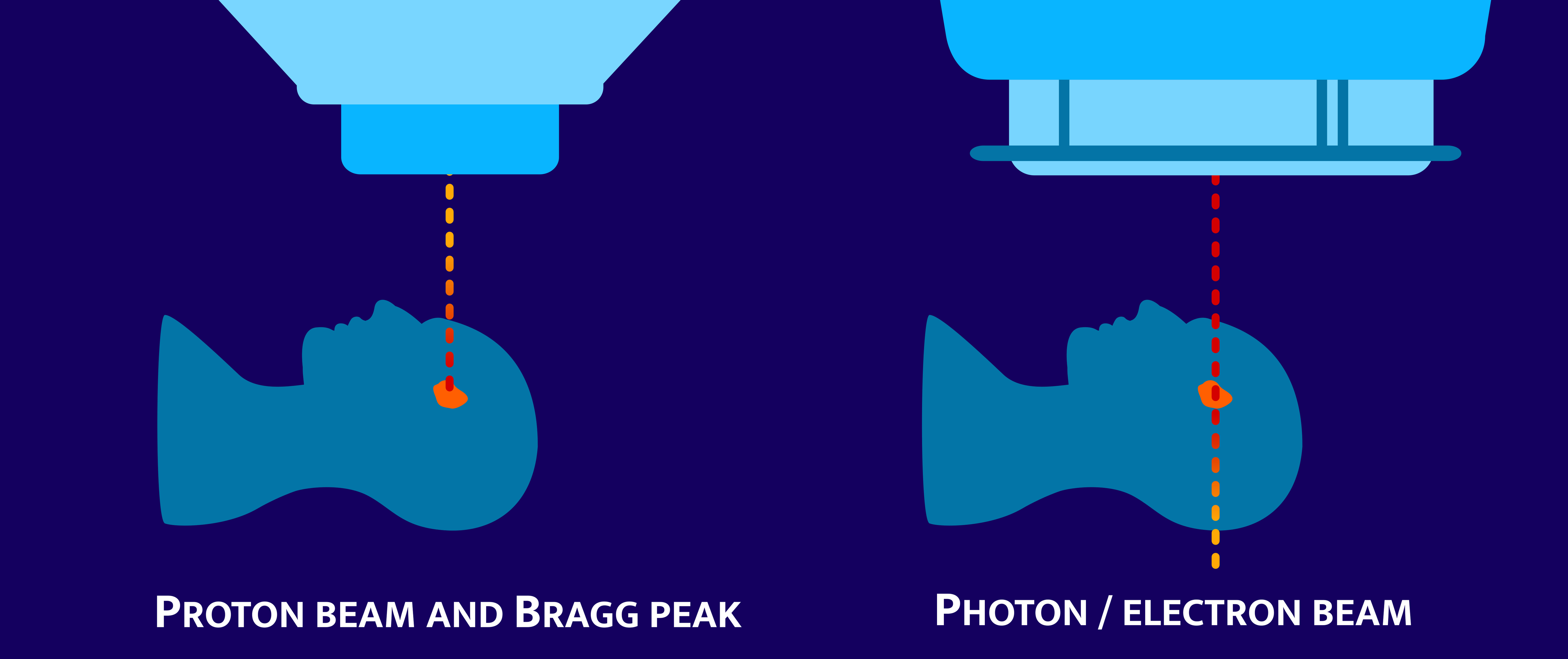
The basic idea of radiotherapy is to deposit a sufficiently large dose of radiation (measured in "Gray") into the tumor without causing lethal damage to healthy tissue. Conventional external radiation therapy uses γ-rays for this purpose, i.e. high-energy X-rays which are emitted by a linear accelerator and then directed at the patient. Unfortunately, these rays not only deposit their dose in the tumor, but a considerable proportion of the dose is also delivered to healthy tissue (see image, right). Young patients in particular run the risk of developing serious late effects after years or decades of therapy. Unfortunately, some tumors do not respond very well to radiation with γ-rays and do not regress sufficiently.
Radiation therapy with protons or carbon ions is intended to help here. Protons (i.e. the atomic nucleus of the hydrogen atom) or carbon atomic nuclei are accelerated to very high speeds using a particle accelerator. At these speeds, these particles develop a special property: they can penetrate tissue and lose very little energy in the process, which means that they emit very little radiation dose to the patient. Only at the end of the path of these particles do they deliver their full dose within a short distance, in the so-called "Bragg peak" (see image, left). In this form of radiotherapy, the aim is therefore to adjust the range of the particles so that they come to a standstill in the tumor and destroy the tumor in the process. It has been found that even tumors that react poorly to γ-rays can still be successfully treated using this form of radiotherapy. Carbon therapy was significantly developed at the Society for Heavy Ion Research in Darmstadt and is used today at the Heidelberg Ion Beam Center Heidelberg Ion Beam Center for patients.
The fact that radiation therapy with protons and carbon ions delivers the dose in a small area makes it particularly important to ensure that the beam is stopped at exactly the right point in the tumor. Despite intensive research and great progress in recent years, it has still not been possible to develop procedures for all cases and patients that enable doctors and clinical staff to monitor the position and range of the ions in the patient with millimeter precision during irradiation.
Our working group is researching new approaches to make it possible in future to monitor the range of the ions during irradiation with the highest precision. We are working closely with the Society for Heavy Ion Research in Darmstadt.